 In keeping with the theme, here's the newest post. Describe what you see. What has changed?
Thursday, December 22, 2011
AMD, what do you see?
In keeping with the theme, here's the newest post. Describe what you see. What has changed?
AMD, Soft Drusen and Geographic Atrophy

Age related macular degeneration (AMD) is the leading cause of central vision loss for Americans age 50 or greater. Risk factors include smoking, intense exposure to sunlight and most importantly, age. Aging of the photoreceptor cells and RPE is thought to be the primary pathophysiologic process contributing to macular degeneration. The two primary types of AMD are "dry" or nonexudative and "wet" or exudative macular degeneration. This post will focus on the previously posted images, highlighting dry AMD.
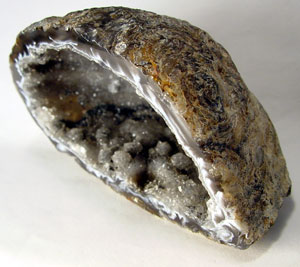
The primary findings in the above images, characteristic of dry AMD, are the cream colored nodules concentrated mostly around the central macular region. These are known as "drusen" which comes from the german word for geode.
These drusen are actually hyaline deposits within Bruch's membrane, which separates the RPE from the choriocapillaris. Drusen may distort the overlying retina enough to cause very subtle visual changes (metamorphopsia), but usually, they remain asymptomatic unless affecting the fovea itself. Drusen may be divided into several categories which represent the pathophysiological progression. Hard drusen are more refractile and have distinct borders. Soft drusen are the cream colored (often larger) bodies with blurred borders. Soft drusen may further coalesce into what are called confluent drusen. While drusen are mostly harmless in and of themselves, they are associated with the visually threatening outcome of dry AMD, Geographic Atrophy (GA). So-called geographic atrophy, when large areas of RPE becomes depigmented (seen as a window defect where the details of the choroid can clearly be seen), is most closely associated with soft and confluent drusen.
Drusen can often be most clearly demonstrated on fluorescein, later posts will highlight these as well as OCT imaging (very important in this day in age).
The BSCS series volume on Vitreoretinal disease devotes a relatively large portion of the text to AMD and is a good source.
Monday, December 19, 2011

Friday, December 16, 2011
What brings you in today? (continued)
 In continuation of yesterday's post, included is the patient's photos from approximately 2 1/2 years before. As was correctly pointed out in the comments, this patient was relying on her left eye for nearly all of her vision. It was the acute change in her better eye that prompted her to notice a change in her vision. The image shows a striking example of the natural progression of AMD, the leading cause of blindness in American adults. The fundus of the right eye is remarkable for the large disciform scar which represents the end stage of choroidal neovascularization (CNV) and wet (or exudative) age related macular degeneration. Now that we live in the era of anti-VEGF injections, advanced disciform scars like this will hopefully be a thing of the past. The left eye was likely still dry in the first set of images. The characteristic finding of advanced dry (nonexudative) AMD that can be seen is the significant geographic atrophy (GA) representing degenerative changes to the pigment epithelium (RPE). Unfortunately, the acute changes resulting in a loss of vision were the result of a hemorrhage heralding the transformation from dry AMD to wet. Stay tuned for more examples of AMD as it is one of the most important disease entities affecting the retina.
Thursday, December 15, 2011
What brings you in today?
 The above images represent a 79 year old caucasian female. She presented to the clinic due to worsening of vision. Which eye prompted this patient to come in for this visit?
Myelenated Nerve Fibers
The finding shown in the previous post represents myelenated nerve fibers (MNF). This is a congenital anomaly that is commonly confused with cotton wool spots (CWS), though does not represent any acute pathology. Usually, MNF are continuous with the optic disc. The key visual clue is the feathery appearance, especially around the margins. Often MNF are asymptomatic (as in this patient), and observation is the only course of action. If the lesion is large enough, or extending into the macula, some degree of scotoma may be present.
Further reading:
http://dro.hs.columbia.edu/myelfibers.htm
http://www.nature.com/eye/journal/v17/n1/full/6700266a.html
Sunday, December 11, 2011

Saturday, December 10, 2011
Carry a stethoscope!
 This case, and the representative images are a wonderful example of how remote illness can affect the eye. It is also a nice reminder that every once in a while, an ophthalmologist may need to dust off the stethoscope.
1) The above color fundus photos of a patient's righ
t eye shows an area of retinal whitening in the temporal region of the macula, sparing the fovea. This whitening represents nerve fiber layer (NFL) ischemia and edema. Hypoxia impairs axonoplasmic flow, which is what leads to the swelling of nerve fibers in this ischemic state. Notice how the fovea remains spared, just as in the "cherry red spot." The image is characteristic of branch retinal artery occlusion (BRAO). The emboli are strikingly seen at approximately the same location along both the superior and inferior temporal arcades. The main clue is that the patient is just now being treated for strep sepsis. From this picture, one must be extremely concerned that an endocarditis has developed and valve vegetations provide the source for these emboli. In fact, we can see that microabscesses have formed at the sites where the bacterial plaque came to rest.
The FA shows the emboli as blockages to arterial filling. Also, areas of diffuse hypoperfusion can be seen corresponding to the area of NFL ischemia.
Here are a couple of views you might see through a direct ophthalmoscope. The top right would represent the "cherry red spot" that would come into view if you asked the patient to fixate on the light. This should prompt you to trace along the arcades to try to find emboli, shown in the other two simulated views.:

2) In the case of any CRAO or BRAO, a stethoscope could be used to listen for carotid bruits or hear murmurs. In this case, the high index of suspicion for septic emboli, a transesophageal ultrasound to look at the heart valves is of the utmost importance. While patients with this picture are usually very sick, it is possible that the first dose of antibiotics has already reduced the bacteremia, thus improving the patient's systemic symptoms.
In this case, the patient was found to have a murmur, and was sent for ultrasound. Aortic valve vegetation was discovered and the patient underwent surgery for replacement of the valve.
More reading:
http://www.ncbi.nlm.nih.gov/pubmed/10409855
http://www.merckmanuals.com/professional/cardiovascular_disorders/endocarditis/infective_endocarditis.html
Sunday, December 4, 2011
Retina and systemic disease. What do you see?

64 year old female with acute visual loss noted one week after starting treatment for strep sepsis.
1)What do you see?
2)What do you want to look at next in the work-up?
Subscribe to:
Comments (Atom)