Sunday, June 2, 2013

Illustrative FAs

This is a 63 year old woman with VA of 20/60 and NLP with poorly controlled DM and lost to follow-up for 2 years.  What are the FA findings, and what can you infer as to the cause of her blindness in the left eye?


Short answer (annotated images to follow):
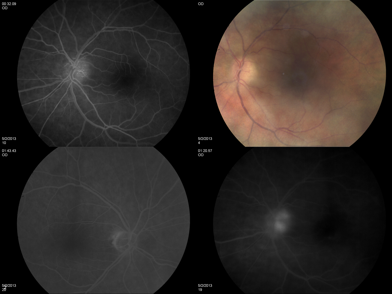
The first image demonstrates a right eye in a patient with PDR. Large areas of non-perfusion can be seen, with adjacent NVE (remember, neovascularization is often found on the border of perfused and non-perfused tissue). The 2nd image is an anterior FA which demonstrates NVI (along with PCIOL, superior iridotomy).  The natural progression of NVI, which can often be seen first at the pupilary border, eventually causes fibrovascular obstruction of the trabecular meshwork, results in neovascular glaucoma.  We can infer that the fellow eye has long since suffered glaucomatous damage to the optic nerve 2/2 NVI, thus resulting in the NLP visual acuity.


Two photos, 4 months apart. What changed?


Saturday, February 23, 2013


This is an 85 year old woman with no visual symptoms. What do you see?

Thursday, February 16, 2012

What do you see?

Describe the above set of images. This represents a leading cause of blindness among working-age Americans.

Your next phone call...

First, my apologies for taking so long between posts. I have been spending long hours on my medicine Sub-I on the Heme/Onc service...which provides a perfect segue into the discussion of the previous image.

This image is a set of color fundus photos showing the left eye. Most remarkable finding in this image are the very prominent hemorrhages. There are both subhyaloid and intraretinal hemorrhages. Several of the intraretinal hemorrhages display white centers, also known by there eponym, "roth's spots."
Roth's spots are often discussed as findings where a patient has septic emboli from subacute bacterial endocarditis (SBE). In reality, though, they are not a specific finding and are associated with many systemic diseases including diabetic retinopathy and various hematologic malignancies.
In addition, this retina shows extremely globular subhyaloid hemorrhages, that show fluid level from the settling of red blood cells. In these subhyaloid areas, there also appears to be large amounts of serous fluid. This picture is very suspicious for bleeding in the setting of severe thrombocytopenia.
In this patient, the acute presentation along with young age, when combined with the sheer extent of pathology raises a red flag for some kind of hematologic process. Key retinal findings are the Roth's spots and macular subhyaloid hemorrhages. In this case, one must quickly rule out an acute leukemia, and a call to a pediatric hematologist/oncologist must be a first priority. The most common pediatric leukemia is Acute lymphoblastic leukemia, which may present in the eye similarly to this patient. http://www.aao.org/theeyeshaveit/optic-fundus/retinal-hemorrhages.cfm
The workup for this patient is ongoing. He was found to have aplastic anemia of unknown (as of yet) etiology. Stay tuned for updates.

Wednesday, January 18, 2012

Your next phone call...

A 16 year old boy presents with sudden decrease of vision. He has been otherwise asymptomatic and even wrestling on the high school team as recently as the past few days. After viewing above, who do you call?

Monday, January 16, 2012

Wet AMD with new subretinal hemorrhage

Here we have an FA and color fundus photo of a left eye at two points in time. The earlier photos are above.

In the color photos, we see some peripapillary pigmentary changes. These are common and, like peripapillary atrophy, crescents, etc, don't amount to much clinically. The primary finding, is the new finding just temporal to the disc. Here we see a subretinal hemorrhage (note the small vessels which pass over the lesion). The FA is ideally read as a dynamic study, and while these images show just one point in time, they are instructive. In the first image, we can see slight hyperfluorescence in the region directly superotemporal to the disc. In the second image, this region is remarkable for a very bright hyperfluorescent lesion. Again, since we aren't given the dynamic series, its not easy to be exact, but what we can say is that the borders of the lesion are indistinct. There is also blocking on the perimeter due to blood. This leasion appears to be leaking, a CNVM, not a static window defect. Final word: juxtapapillary choroidal neovascular membrane.
Juxtapapillary CNVM represents a form of extrafoveal CNVM in wet AMD. Just as in all wet AMD, the CNVM occurs as a result of a disruption in Bruch's membrane which allows for abnormal growth of choroidal vessels which invade the subretinal and intraretinal space. These vessels are leaky and fragile. Leaking of fluid leads to the distortion of the neurosensory retinal architecture. Fragile vessels often break leading to hemorrhage as seen here.
Historically, CNVM has been described as either "classic" or "occult" and may have elements of both forms. Classic CNVM is seen as having distinct margins on FA, whereas occult often is patchy and shows stippled, irregular leakage of FA. Stay tuned for more examples in future image cases.


Thursday, January 5, 2012

First Case of 2012


What do you see?

Progression of GA in Dry AMD


Finally returning from a long holiday break. Happy new year to everybody! Now back to business...

The previous case provides a good example of the progression of geographic atrophy (GA) in dry AMD. We see a set of color fundus photos and FA taken approximately 1.75 years apart. The discs look grossly normal with no significant cupping. There is some peripapillary atrophy, that is not out of the ordinary if seen on its own. The vessels look grossly normal. What we see in the macular region are areas of RPE atrophy which appear as areas where the choroidal details are more distinct. Notice, also, areas of hyperpigmentation along the margin of atrophic areas. As the atrophy progresses in the later set of images, the region becomes more distinct, larger, and more delineated. Note that the ideal imaging modality to evaluate GA is fundus auto-fluorescence, which would clearly demonstrate a rim of hyperfluorescent RPE surrounding black regions of atrophy (non-fluorescent, since RPE is absent).

Dry AMD may progress to an advanced form in a minority of patients. GA represents this advanced form. GA tends to occur in older patients than those with neovascular or wet AMD at the time of presentation. GA is closely associated with pigmentary changes and drusen within the macula. The development of GA is especially linked to large soft drusen. Large, confluent drusen over 125 microns are considered a risk factor for progression to GA. There is often a high degree of symmetry between fellow eyes. Peripapillary atrophy is very common in eyes with GA. The precise pathophysiologic mechanism for the progression of GA, which affects the RPE, choriocapillaris and neurosensory retina is poorly understood.

(Laud K, Mukkamala SK, Brue C, Slakter J. "The Future of Non-neovascular Age-related Macular Degeneration." Ho AC, Regillo CD (eds.) Age-related Macular Degeneration Diagnosis and Treatment, Springer Science+Business Media, LLC. 2011)

Thursday, December 22, 2011

AMD, what do you see?

In keeping with the theme, here's the newest post. Describe what you see. What has changed?

AMD, Soft Drusen and Geographic Atrophy


Age related macular degeneration (AMD) is the leading cause of central vision loss for Americans age 50 or greater. Risk factors include smoking, intense exposure to sunlight and most importantly, age. Aging of the photoreceptor cells and RPE is thought to be the primary pathophysiologic process contributing to macular degeneration. The two primary types of AMD are "dry" or nonexudative and "wet" or exudative macular degeneration. This post will focus on the previously posted images, highlighting dry AMD.

The primary findings in the above images, characteristic of dry AMD, are the cream colored nodules concentrated mostly around the central macular region. These are known as "drusen" which comes from the german word for geode.
These drusen are actually hyaline deposits within Bruch's membrane, which separates the RPE from the choriocapillaris. Drusen may distort the overlying retina enough to cause very subtle visual changes (metamorphopsia), but usually, they remain asymptomatic unless affecting the fovea itself. Drusen may be divided into several categories which represent the pathophysiological progression. Hard drusen are more refractile and have distinct borders. Soft drusen are the cream colored (often larger) bodies with blurred borders. Soft drusen may further coalesce into what are called confluent drusen. While drusen are mostly harmless in and of themselves, they are associated with the visually threatening outcome of dry AMD, Geographic Atrophy (GA). So-called geographic atrophy, when large areas of RPE becomes depigmented (seen as a window defect where the details of the choroid can clearly be seen), is most closely associated with soft and confluent drusen.

Drusen can often be most clearly demonstrated on fluorescein, later posts will highlight these as well as OCT imaging (very important in this day in age).

The BSCS series volume on Vitreoretinal disease devotes a relatively large portion of the text to AMD and is a good source.


Monday, December 19, 2011

What do you see?


What is going on in the macula? This is a risk factor for what?

Friday, December 16, 2011

What brings you in today? (continued)

In continuation of yesterday's post, included is the patient's photos from approximately 2 1/2 years before. As was correctly pointed out in the comments, this patient was relying on her left eye for nearly all of her vision. It was the acute change in her better eye that prompted her to notice a change in her vision. The image shows a striking example of the natural progression of AMD, the leading cause of blindness in American adults. The fundus of the right eye is remarkable for the large disciform scar which represents the end stage of choroidal neovascularization (CNV) and wet (or exudative) age related macular degeneration. Now that we live in the era of anti-VEGF injections, advanced disciform scars like this will hopefully be a thing of the past. The left eye was likely still dry in the first set of images. The characteristic finding of advanced dry (nonexudative) AMD that can be seen is the significant geographic atrophy (GA) representing degenerative changes to the pigment epithelium (RPE). Unfortunately, the acute changes resulting in a loss of vision were the result of a hemorrhage heralding the transformation from dry AMD to wet. Stay tuned for more examples of AMD as it is one of the most important disease entities affecting the retina.